Etiology: Escherichia coli are Gram-negative bacilli. E. coli recovered from hamsters with colibacillosis has virulence factors that allow for adherence to mucosal cells.
Incidence: The incidence of disease is rare in research animals. Certain strains of hamsters may be more susceptible than others, i.e., long-haired fawn and teddy bear.
Transmission: The disease is transmitted via direct contact, fecal-oral and fomite contamination. A high carbohydrate or vitamin imbalanced diet, exposure to infected animals, shipment stress, overcrowding, and lack of fresh water are stressful events that precipitate development of disease.
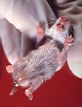
Clinical Signs: Generally the disease is acute in onset, with 2- to 4-week-old hamsters developing a profuse yellow watery diarrhea that mats the area around the tail. Dehydration and death quickly ensues.
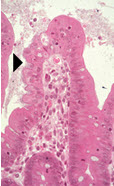
Pathology: Lesions are most obvious in the small intestine and cecum. Fluid and gas dilate the small intestine and cecum, occasional serosal edema is observed. The stomach is usually empty and gas-filled. There is submucosal edema and effacement of enterocytes colonized by bacteria in the small intestine or cecum (arrowhead). Often, there is little to no inflammatory cell infiltrate in the lamina propria or submucosa.
Diagnosis: Culture of the gut with recovery of pure culture of E. coli is strong evidence for disease. Evaluation for virulence factors, such as the production of toxins, or of proteins necessary for adherence and/or invasion is required to differentiate the isolate from the nonpathogenic E. coli that may be found in low numbers in healthy hamster gut.