Etiology: LCMV is an enveloped RNA virus of the arenavirus group.
Incidence: The natural reservoir for LCMV is the wild rodent population. Incidence of infection and spontaneous disease is rare. Most reported human cases have been associated with infected pet hamsters.
Transmission: Transmission occurs via urine and saliva, traumatized skin, conjunctiva, respiratory passages, or congenital contamination. If infected as young adults, antibody production and persistent viruria and viremia continue for up to 6 months. In research involving hamsters, another route of infection could be from the transplantation of LCMV-contaminated tumors.
Clinical Signs: Usually there are no clinical signs. In utero or perinatal infections (within 1 day post-partum) may produce a subclinical persistent infection or a chronic, progressive wasting disease. Signs can include convulsions, decreased growth, and inactivity. Decreased reproduction has been reported in chronically infected females.
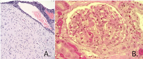
Pathology: Gross lesions vary. If present, lesions may include splenomegaly, swollen or shrunken, pitted kidneys, lymphadenopathy and hepatomegaly. Microscopic lesions include lymphocytic meningitis (A.), chronic glomerulonephropathy (B.), widespread vasculitis, and marked lymphocytic infiltration of the viscera.
Diagnosis: Diagnosis is based partly upon histologic observation of lymphocytic infiltration of the meninges, choroid plexus, and of submeningeal and subchoroid perivascular spaces. MFI or IFA tests can be used but may be of limited value in endemically infected animals. PCR of hamster tissue or transplantable cells/fluids should be used to diagnose persistent or acute infections.
Public Health Significance: People are susceptible to LCMV, and experience flu-like symptoms and occasional nonsuppurative meningitis. Past reports of zoonosis have linked human infections to exposure to infected pet hamsters [1].