Etiology: Syphacia mesocricetus is the hamster pinworm. Syphacia obvelata and Syphacia muris are also capable of infecting hamsters.
Incidence: Prevalence of a pinworm infection is low, however, the incidence of parasitism within individual colonies may be high.
Transmission: Transmission occurs via ova ingestion. The eggs are very light and have been shown to aerosolize, resulting in widespread exposure. Syphacia species adult females migrate from the cecum through the colon to the rectum, and deposit their eggs in a bolus on the perianal area. Eggs of Syphacia species are infective within 5-20 hours.
Distribution: Adult Syphacia species are found in the cecum.
Clinical signs: No signs are usually seen. Heavy parasite loads may lead to rectal prolapse or perianal irritation.
Diagnosis:
Antemortem: Cellophane tape test of the perianal area should be used to detect ova of Syphacia species.
Postmortem: Place opened cecum and colon in a Petri dish containing saline. In a short amount of time, the pinworms will migrate out of the gut lumen into the saline. The pinworms can be detected with a dissecting microscope, and speciated with use of light microscopy.
Diagnostic Morphology:
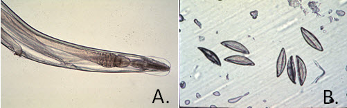
Syphacia mesocricetus: Round esophageal bulb. Broad cervical alae, ending abruptly 1/2 way to the esophageal bulb (A).
Female: 3.2-6.9 mm in length. Vulva in anterior 1/8 of body (700-800 µm from anterior end).
Male: 1.1-1.5 mm in length. Tail is long and pointed. Three prominent ventral mammelons; center mammelon near the middle of the body lengthwise.
Ova: 130-140 x 40-50 µm; pseudo-operculated, thin-shelled (B).