Etiology: This disease is similar to proliferative bowel disease in swine and ferrets. In swine, increased understanding of biochemical and physical structures of the organisms has led to the classification of the bacillus as Lawsonia intracellularis. L. intracellularis is also believed to be the etiologic agent associated with proliferative ileitis in hamsters [2]. The literature also identifies other possible causative or contributive agents such as lactose negative E. coli, Campylobacter coli, and chlamydial organisms.
Incidence: The incidence of this disease is rare.
Transmission: The disease is transmitted via direct fecal-oral contact and fomite contamination.
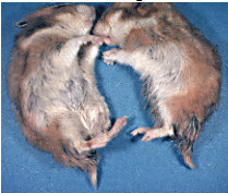
Clinical Signs: Acute disease is manifest by lethargy, anorexia, irritability, ruffled hair, diarrhea, dehydration, and death. The moist feces stain the base of the tail. The disease occurs primarily in hamsters 3 to 10 weeks of age, has been noted in adult animals debilitated by concurrent disease(s).
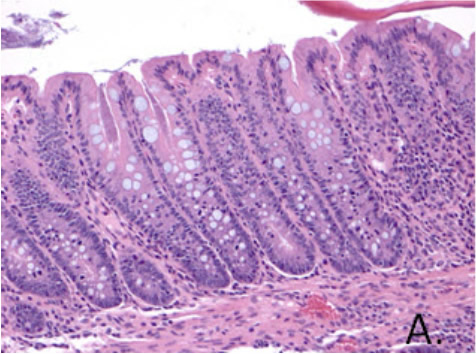
Pathology: Lesions are most obvious in the ileum and cecum. Fluid feces dilate the ileum and cecum, and edema and hemorrhage may be observed on the serosa. The ileum may appear more rigid (tubular) and thicker on cross-section. On histologic exam, affected intestinal segments are affected with mild to moderate segmental hyperplasia of the mucosa. The crypts are elongated and tortuous with marked proliferation of enterocytes and occasionally goblet cells (A.).
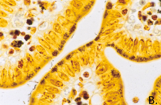
Lymphoid hyperplasia and edema are often present. Migrating leukocytes are seen near the base of the villi. The villous tips undergo coagulation necrosis as the disease progresses. Additional bacterial involvement may cause accumulation of purulent exudate in the crypts and formation of microabscesses in the intestinal wall. Silver stains (B.) are needed to reveal numerous curved bacilli in the apical cytoplasm of the enterocytes.
Diagnosis: Diagnosis is made primarily by recognition of characteristic gross and histologic lesions. Verification of the causative agent can be accomplished by a polymerase chain reaction (PCR) assay using fresh or frozen gut samples.
2. The Laboratory Rabbit, Guinea Pig, Hamster, And Other Rodents. 1 ed2012, 225 Wyman Street, Waltham, MA 02451: Elsevier.