Etiology: Pneumocystis is a single-celled fungal (Ascomycota) respiratory pathogen of mammals. P. carinii and P. wakefieldiae are the two Pneumocystis species that have been identified in laboratory rats, with P. carinii being most commonly found.
Incidence: The incidence of infection is common.
Transmission: Transmission occurs via direct contact and airborne routes. Experimental infection studies have shown that Pneumocystis isolates are host species-specific. Interspecies transmission does not occur even in immunodeficient hosts.
Clinical Signs: Immunodeficient rats with pneumocystosis present with weight loss, ruffled fur or dry skin and a hunched posture. Pneumocystis is known to cause a severe, often lethal pneumonia in immunocompromised hosts. Neonatal rats which acquire infection within hours after birth and can harbor the organism without evidence of clinical disease unless subjected to chronic immune suppression.P. carinii has been found in the lungs of clinically healthy commercially produced immunocompetent rats.
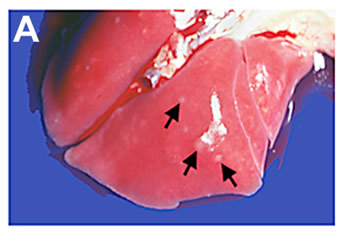
Pathology: Gross pulmonary lesions include 1-4 mm grey, flat to raised foci randomly distributed throughout all lung lobes at the peak of disease expression (arrows, A.).
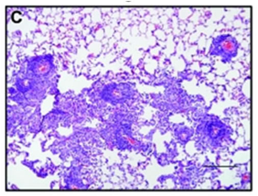
Lesion severity is greatest at 5 wk after exposure and consists of moderate to severe multifocal perivascular lymphohistiocytic infiltrates and moderate to severe multifocal lymphohistiocytic interstitial pneumonia. In areas of interstitial pneumonia, there is occasional to marked type 2 pneumocyte hyperplasia, and many alveolar spaces are filled with macrophages (C) [1].
Diagnosis: Diagnosis is made by performing PCR of lung tissue, serology in immunocompetent animals and histopathologic survey of lung tissue.
1. Livingston, R.S., et al., Pneumocystis carinii infection causes lung lesions historically attributed to rat respiratory virus. Comp Med, 2011. 61(1): p. 45-59.